


"Exploring the Essentials of Sciatic Nerve Anatomy"

For those grappling with the challenges of sciatica, I´ll offer a brief explanation of the anatomy of the sciatic nerve.
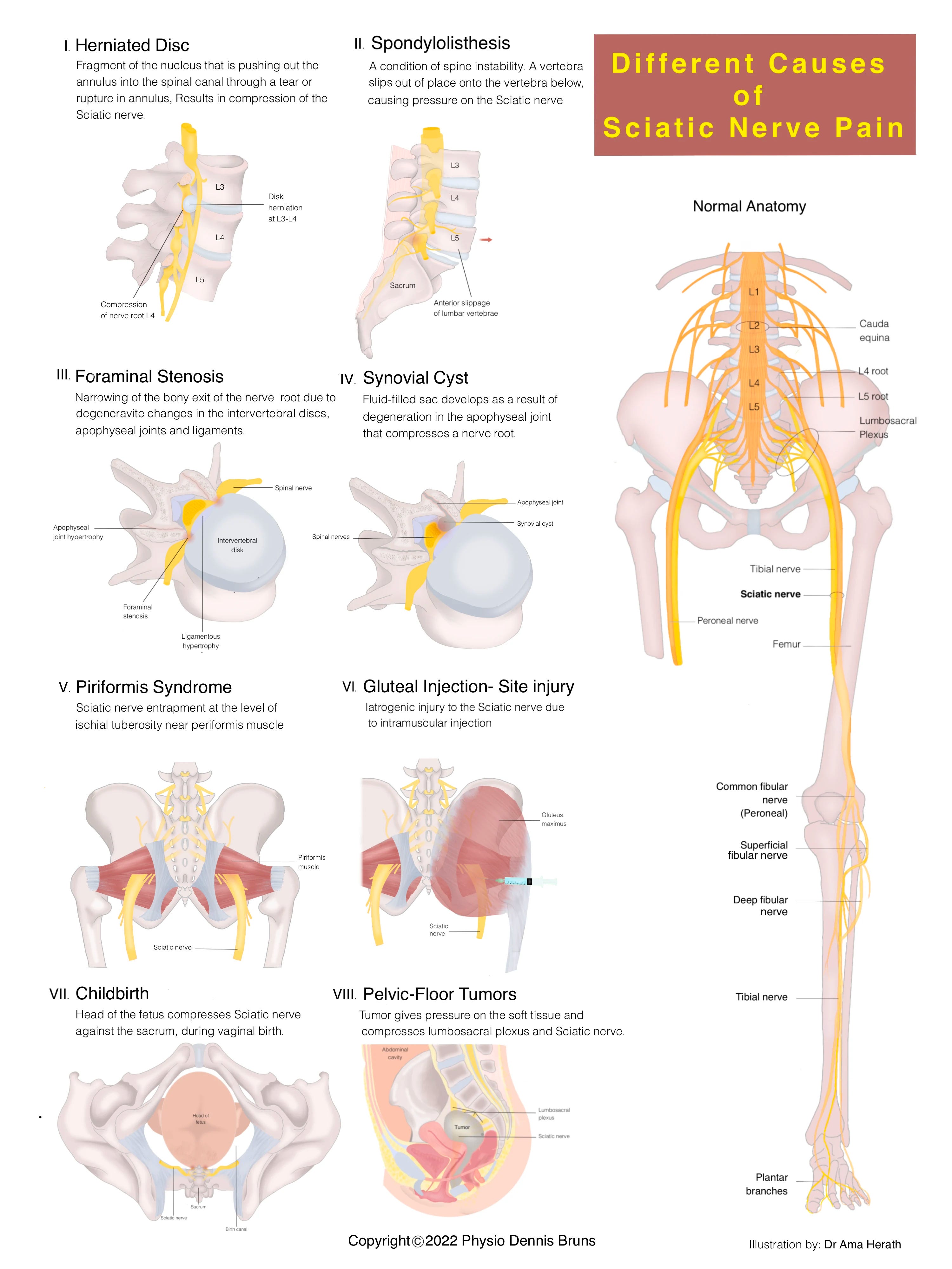
The sciatic nerve is the body's largest nerve, spanning from the lower back all the way down to the heel of the foot. Its significance lies in its responsibility for both controlling and conveying sensations from a substantial portion of the muscles and skin in the thigh, leg, and foot.
This formidable nerve originates from a convergence of spinal nerves in the lower back, unifying into a singular, robust nerve upon leaving the spine. As it departs the spine, it traverses through various pelvic structures and muscles, eventually descending down the posterior aspect of the thigh.
As it approaches the knee, the sciatic nerve diverges into two critical branches:
Tibial Nerve: This branch proceeds along the posterior aspect of the leg and foot.
Common Peroneal Nerve: This counterpart takes a route to the anterior and lateral aspects of the leg and foot.
Consider the sciatic nerve as the central command for your leg muscles. It orchestrates the functioning of thigh muscles necessary for knee flexion and hip movements. The tibial nerve plays a pivotal role in activating calf muscles and facilitating foot movements, while the common peroneal nerve oversees the muscles responsible for foot dorsiflexion and toe extension.
In addition to its motor functions, the sciatic nerve serves as a sensory conduit, transmitting signals from the leg and foot to the brain. It's worth noting that the inner leg relies on a separate nerve for sensation. The tibial nerve, in particular, assumes the specialized role of conveying sensory information from the sole of the foot.
To maintain its health and functionality, the sciatic nerve boasts its own dedicated blood supply, sourced from neighboring arteries and veins.
The sciatic nerve's structure and path can vary. Typically, it exits below the piriformis muscle. However, it can also divide into two parts above the piriformis, leading to six variations.
*
Contact the author for more information:
-www.youtube.com/@physiodennisbruns
-www.tiktok.com/@physiodennisbruns
*
References:
Atoni AD, Oyinbo CA, Francis DAU, Tabowei UL. Anatomic Variation of the Sciatic Nerve: A Study on the Prevalence, and Bifurcation Loci in Relation to the Piriformis and Popliteal Fossa. Acta Med Acad. 2022 Apr;51(1):52-58.
Bartret AL, Beaulieu CF, Lutz AM. Is it painful to be different? Sciatic nerve anatomical variants on MRI and their relationship to piriformis syndrome. Eur Radiol. 2018 Nov;28(11):4681-4686.
Bharadwaj UU, Varenika V, Carson W, Villanueva-Meyer J, Ammanuel S, Bucknor M, Robbins NM, Douglas V, Chin CT. Variant Sciatic Nerve Anatomy in Relation to the Piriformis Muscle on Magnetic Resonance Neurography: A Potential Etiology for Extraspinal Sciatica. Tomography. 2023 Feb 22;9(2):475-484.
Poutoglidou F, Piagkou M, Totlis T, Tzika M, Natsis K. Sciatic Nerve Variants and the Piriformis Muscle: A Systematic Review and Meta-Analysis. Cureus. 2020 Nov 17;12(11):e11531.
Reynoso JP, De Jesus Encarnacion M, Nurmukhametov R, Melchenko D, Efe IE, Goncharov E, Taveras AA, Ramirez Pena IJ, Montemurro N. Anatomical Variations of the Sciatic Nerve Exit from the Pelvis and Its Relationship with the Piriformis Muscle: A Cadaveric Study. Neurol Int. 2022 Oct 31;14(4):894-902.
Robinson DR. Pyriformis syndrome in relation to sciatic pain. Am J Surg. 1947 Mar;73(3):355-8.
Ro TH, Edmonds L. Diagnosis and Management of Piriformis Syndrome: A Rare Anatomic Variant Analyzed by Magnetic Resonance Imaging. J Clin Imaging Sci. 2018;8:6.